Users Currently Browsing This Topic:
0 Members

Author Topic: 11/22/63 Parkland Medical Reports, the Throat Wound, and the Large Head Wound  (Read 10964 times)

Offline Michael T. Griffith

  • Hero Member
  • *****
  • Posts: 929
Advertisement
Let us continue to compare the Parkland descriptions of JFK’s large head wound with the Bethesda descriptions.

Keep in mind that part of the temporal bone is behind the ear, and that its lower half borders the occiput. The occiput is the main bone in the back of the head. Most of the parietal bone is behind the ear, and the lower part of the parietal bone borders the temporal and occipital bones.

Dr. John Ebersole, the radiologist at the autopsy

Dr. John Ebersole was the radiologist at the autopsy. He directed the taking of the autopsy x-rays. He was interviewed by the HSCA forensic pathology panel (FPP). Dr. Ebersole said that “the back of the head was missing” (HSCA interview transcript, 3/11/1978, p. 3). He added that he “personally held the head” (HSCA interview transcript, 3/11/1978, p. 10).

When the FPP showed Dr. Ebersole autopsy photos F3 (back of the head) and F5 (lower part of the back of the head) and asked him about the large head wound, he said that the photos showed the wound much farther forward and lateral then he remembered seeing it, “much more lateral and superior [forward] than I remembered,” and that ”my recollection is more of a gaping occipital wound than this.... I would have put the gaping wound here rather than more forward” (HSCA interview transcript, 3/11/1978, pp. 62-63).

By the way, Dr. Ebersole reported that the autopsy doctors learned of the throat wound during the autopsy, just as Dr. Malcolm Perry said, and contrary to Dr. Humes’s lie that he knew nothing about it until the next morning (HSCA interview transcript, 3/11/1978, pp. 4-5). Dr. Ebersole also reported that “a fragment of the occipital bone” (i.e., bone from the back of the head) arrived late during the autopsy, and that he x-rayed it (HSCA interview transcript, 3/11/1978, p. 5).

James Curtis Jenkins, a lab technician at the autopsy

James Jenkins was a lab technician at Bethesda Naval Hospital and saw part of the autopsy.

Jenkins told the HSCA that the large head wound was in the "middle temporal region back to the occipital" (HSCA interview transcript, 8/29/1977, p. 4). The middle temporal region back to the occiput is in the right-rear part of the head.

In a recorded interview with JFK research William Matson Law, Jenkins said,

"You could see that the area where the bone was actually missing was in the parietal-occipital area in the back of the head and extended downward touching the temporal area." (Jenkins and Law, At the Cold Shoulder of History, Trine Day, 2018, pp. 31-32)

Edward Reed, x-ray technician at the autopsy

Edward Reed was one of the x-ray technicians at the autopsy. He told the HSCA that the large head wound was “located in the right hemisphere in the occipital region” (HSCA interview transcript, 5/2/1978, p. 2).

There were three Bethesda witnesses who changed their descriptions of the large head wound when they were interviewed by the ARRB: Edward Reed, Jerrol Custer, and John Stringer. Fortunately for the sake of accurate history, all three had given recorded interviews years earlier in which they specified that the large head wound was in the back of the head. When they changed their tune with the ARRB, the ARRB interviewers confronted them with their previous statements. Reed and Custer would not budge even after being confronted with their earlier statements, but Stringer, after being played a recording of him saying the wound was in the back of the head, was eventually willing to acknowledge that there was bone missing from the occiput.

Someone appears to have rehearsed Reed’s testimony with him before his ARRB interview and told him what to say. Why? Because Reed claimed that he saw the 6.5 mm object on the AP x-ray on the night of the autopsy. Reed was the only Bethesda witness who said he saw the 6.5 mm object on an x-ray at the autopsy. Every other Bethesda witness, including all three autopsy doctors, who was asked about the 6.5 mm object said they did not see it on any x-ray that night. And, of course, we now know that the 6.5 mm object is a ghosted image that was added to the AP x-ray after the autopsy. So Reed was clearly lying to the ARRB, whereas his HSCA testimony about the large head wound agrees with that of the vast majority of the witnesses who saw the wound.

John Stringer, medical photographer at the autopsy

John Stringer took most of the autopsy photos. When interviewed, on tape, in 1972, he said the large head wound was in the back of the head:

LIFTON: Was the main damage to the skull on the top, or in the back?
STRINGER: In the back.
LIFTON: High In the back, or lower In the back?
STRINGER: Oh, the occipital part in the back there (garbled) up above the neck.
LIFTON: In other words, the main part of his head that was blasted away was in the occipital part of the skull?
STRINGER: Yes. the back part. (https://www.history-matters.com/essays/jfkmed/TracesOfWitnessTampering/TracesOfWitnessTampering.htm; see also ARRB interview transcript, 7/16/1996, pp. 76-77. The ARRB played the recording of the 1972 interview for Jenkins during his deposition.)

In his ARRB deposition, Stringer initially described the large head wound as being mainly parietal and above and forward of the right ear. Then, the ARRB interviewer played the above-quoted portion from Stringer’s 1972 interview. Under what can be called cross-examination, Stringer said that there was in fact bone missing from the occiput and that the occiput was fractured; in referring to the occiput, he added, “some of the bone was still there,” which of course means that some of it was not (ARRB interview transcript, 7/16/1996, pp. 89-90).

Floyd Riebe, medical photographer at the autopsy

Floyd Riebe was a medical photographer at the autopsy; he assisted John Stringer.

Riebe told the HSCA that he saw a “large wound located around the rear of the head near the top” (HSCA interview transcript, 5/12/1978).

In the first part of his ARRB deposition, Riebe described the large head wound as being “occipital” and on the “right side of the back” of the head, and he even put his hand over the back of his head to demonstrate (ARRB interview transcript, 5/17/1997, pp. 44-45).

But, then, later in the interview, when shown autopsy photos F3 and F5, Riebe did a remarkable about-face and said he must have been mistaken about the wound’s location (ARRB interview transcript, 5/17/1997, p. 71). When pressed to explain his marked change of testimony, Riebe blamed the change on the “chaos” during the autopsy and claimed he must have just “misjudged” where the wounds were (ARRB interview transcript, 5/17/1997, p. 77).

This does not sound believable. Riebe was a trained medical photographer who had spent time at the Armed Forces Institute of Pathology. He surely knew the difference between a wound in the occipital region and a wound above and forward of the right ear in the parietal region, regardless of how much “chaos” there was at the autopsy. During his HSCA interview 19 years earlier, Riebe expressed no doubt about the wound’s location.

Furthermore, when Riebe was shown the autopsy photos of the head years earlier, he insisted they did not show the wounds that he saw, saying, "The two pictures you showed me are not what I saw that night." When asked to describe the large wound, Riebe held his hand over the back of his head and said, "It had a big hole in it. This whole area was gone." With regard to the pictures and X-rays, Riebe said, "It's being phonied some place. It's make believe” (http://jfk.hood.edu/Collection/Weisberg%20Subject%20Index%20Files/L%20Disk/Livingstone%20Harrison%20Edward/Item%20072.pdf, pp. 8-9).

Dennis David, “chief of the day” at Bethesda on the day of the autopsy

David was a senior non-commissioned officer stationed at Bethesda. He was “chief of the day” at the hospital on the day of the assassination. He was at the morgue when JFK’s body arrived at 6:35 PM. He saw some of the autopsy photos a few days after the autopsy. A good friend of his, William Pitzer, was in charge of the Bethesda Naval Hospital’s audio-visual department, and Pitzer showed him some of the autopsy photos.

David told the ARRB that some of the photos “clearly showed a gaping wound in the back of the President's head and that the top of the head looked intact” (ARRB interview transcript, 2/6/1997, p. 4).

Dr. Robert Karnei, resident doctor at Bethesda

Dr. Robert Karnei was a second-year resident at Bethesda. He was on duty the day of the assassination and saw parts of the autopsy from a distance in the morgue. He left the morgue several times during the autopsy to perform various duties.

Dr. Karnei told the ARRB that he saw two wounds on JFK’s head, one above the right ear and the other “in the posterior skull, up high in the back of the head”; he added that the posterior wound was either in the center or just off-center, and that the “upper posterior skull sagged a bit” (ARRB interview transcript, 5/21/1996, pp. 2-3).

Given that Dr. Karnei did not see the head or body up-close, and given that he said the wound was in the upper part of the back of the head, the posterior wound he described must have been the same posterior wound described by Robinson, Hagan, Spencer, O’Donnell, etc. It could not have been the rear head entry wound because that wound was just above the EOP, and because we now know there was no entry wound in the cowlick.

Dr. Robert Canada, commanding officer of Bethesda treatment hospital

Dr. Robert O. Canada was the commanding officer of the treatment hospital at Bethesda Naval Hospital. He witnessed the autopsy.

Dr. Canada told historian Michael Kurtz in 1968 that he saw a “very large, 3-5 cm wound in the right rear of the President’s head, in the lower right occipital region,” and that the wound was “clearly an exit wound” because the occipital bone was “avulsed” (blown outward) (Kurtz, The JFK Assassination Debates: Lone Gunman versus Conspiracy, University Press of Kansas, 2006, p. 51; see also https://www.fff.org/explore-freedom/article/altered-history-exposing-deciet-and-deception-in-the-jfk-assassination-medical-evidence-part-1/, segment on Dr. Canada begins at 1:08:20). Dr. Canada asked Dr. Kurtz not to reveal his account until 25 years after he died, so Kurtz did not write about until 2006.

Incidentally, Dr. Canada also said that the back wound was at around “T3,” that the bullet “did not exit,” and that its wound tract ended in the chest near the stomach (Kurtz, The JFK Assassination Debates, p. 91).

Paul O’Connor, medical technician at the autopsy

Paul O’Connor was a Navy medical technician at Bethesda who assisted with autopsies and who usually helped to perform autopsy craniotomies. He helped remove JFK’s body from the casket and witnessed much of the autopsy; he also saw part of the burial-preparation work done by the Gawler’s Funeral Home technicians.

O’Connor told the HSCA that the large head wound was in the “occipital around the temporal and parietal regions,” and that it appeared to him that the bullet “came in from the right front” (HSCA interview transcript, 8/29/1977, pp. 5-6).

When the HSCA interviewer showed O’Connor CE 386, the Rydberg medical illustration of the head wounds done for the WC, O’Connor said the drawing “did not reflect what I saw” (HSCA interview transcript, 8/29/1977, p. 9).

When interviewed in 2003, O’Connor said that the autopsy photo of the back of the head was false, and that the back of the head was “blown away”:

“One picture of the back of his head shows a complete skull and the hair is untouched,” said O’Connor, who as a 22-year-old Navy corpsman at Bethesda Naval Hospital in Maryland assisted in the autopsy of the assassinated president. “But it was all blown away.” (https://www.gainesville.com/article/LK/20031122/news/604167677/GS)

Interestingly, and in agreement with Robinson and VonHoesen, O’Connor told the HSCA that part of the large head wound had to be filled with rubber and cement because there was still bone missing from it even after the skull fragments from Dallas arrived (HSCA interview transcript, 8/29/1977, p. 7).
« Last Edit: August 14, 2020, 12:35:55 AM by Michael T. Griffith »

JFK Assassination Forum


Offline John Mytton

  • Hero Member
  • *****
  • Posts: 4267
This spurious argument was debunked decades ago. Let us start by quoting from Dr. McClelland's WC testimony:

Mr. SPECTER. Before proceeding to describe what you did in connection with the tracheostomy, will you more fully describe your observation with respect to the head wound?
Dr. McCLELLAND. As I took the position at the head of the table that I have already described, to help out with the tracheotomy, I was in such a position that I could very closely examine the head wound, and I noted that the right posterior portion of the skull had been extremely blasted. It had been shattered, apparently, by the force of the shot so that the parietal bone was protruded up through the scalp and seemed to be fractured almost along its right posterior half, as well as some of the occipital bone being fractured in its lateral half, and this sprung open the bones that I mentioned in such a way that you could actually look down into the skull cavity itself and see that probably a third or so, at least, of the brain tissue, posterior cerebral tissue and some of the cerebellar tissue had been blasted out. (2 H 33)


Quote
this sprung open the bones that I mentioned in such a way that you could actually look down into the skull cavity itself

Thanks for posting McClelland's own words, now explain how McClelland could look down into a wound on the back of Kennedy's head and see a third of the brain missing, how could he even make such a declaration if there was only a hole on the back of Kennedy's head? Whereas the official wound location as verified in the Zapruder film and the officially authenticated Autopsy photos is the only logical explanation for this telling observation.


JohnM
« Last Edit: August 14, 2020, 02:40:46 AM by John Mytton »

Offline Michael T. Griffith

  • Hero Member
  • *****
  • Posts: 929
Thanks for posting McClelland's own words, now explain how McClelland could look down into a wound on the back of Kennedy's head and see a third of the brain missing, how could he even make such a declaration if there was only a hole on the back of Kennedy's head?

So your "answer" to all the mutually corroborating Bethesda and Parkland descriptions of the large head wound is to nit-pick McClelland's verbiage and to once again post your goofy top-of-head GIF? That's it?
Oh, boy.

So you're going to latch onto a hyper-literal interpretation of "look down" and simply ignore the fact that McClelland's description of the large head wound closely corresponds to nearly all the other descriptions of the wound? It is amazing to see the difference in how you treat witnesses like McClelland and how you treat witnesses like Brennan.

Anyway, when Nurse Hutton applied a pressure dressing to the back-of-the-head wound (she specified it was in the back of the head), she would have at least slightly lifted the head to position the dressing, and McClelland, if nothing else, could have easily seen the wound and seen the dressing applied over the wound.

McClelland's description of the large head wound closely agrees with the descriptions in the 11/22/63 Parkland medical reports, which were written just hours after JFK died. His description and drawing exactly match Clint Hill's description of the wound, and Hill saw the wound for several minutes up-close in the limo and saw the same wound again at Bethesda. McClelland's description is also very similar to the descriptions given by Nurse Bowron, who packed the large head wound with gauze squares, and by Tom Robinson, the mortician who reassembled JFK's skull after the autopsy and who put rubber in the orange-sized hole in "the back of the head."


Whereas the official wound location as verified in the Zapruder film and the officially authenticated Autopsy photos is the only logical explanation for this telling observation. JohnM

You know this is false. I already refuted this claim, but you simply keep ignoring the refutation and keep repeating the claim. The "officially authenticated autopsy photos" do not even agree with themselves. F8 shows a very different back of the head than F3 and F5 show, and OD measurements indicate there is occipital bone missing in the area where the manmade white patch was placed on the lateral skull x-rays. Are you ever going to deal with the hard scientific evidence that the white patch must be manmade? Why do you suppose that patch was placed there?

By the way, your "official authentication" was done by the same folks--the HSCA FPP--who mislocated the rear head entry wound by 4 inches, who "identified" a rear entry wound in the cowlick that is not seen on the skull x-rays, who erroneously said that the upper-skull fractures radiate from their proposed cowlick entry site when in fact they do not (which also refutes their cowlick entry site), who said the 6.5 mm object was a bullet fragment, who published altered copies of the skull x-rays to conceal the clear evidence of a frontal-shot cluster of fragments in the front part of the skull, etc., etc. Wow, that's some "authentication."

The FPP's report falsely claimed that the Bethesda descriptions of the large head wound agreed with the autopsy report and disagreed with the Parkland descriptions, a claim that we now know is false. The HSCA sealed all the Bethesda medical interviews, and when the ARRB released those interviews, we learned that the Bethesda descriptions overwhelmingly agree with the Parkland descriptions. We also learned that one of the FPP staffers objected to the report's false claim about the Bethesda descriptions, but the authors of the FPP report (Loquvam and Weston) ignored his objection.

When are you going to beam yourself into at least the mid-2000s and start dealing with the scientific and research developments that began to occur in the case in the mid-1990s? You keep pretending that the HSCA medical interviews have not been released, that three medical doctors have not independently done OD measurements on the skull x-rays, that OD measurements have not proved beyond all doubt that the right-rear white patch on the lateral skull x-rays is manmade, that the Zapruder film was not diverted to two CIA photo labs, that F8's orientation has not been conclusively established, that F8 does not show substantial bone missing from the occiput, that the Harper fragment has not been established as being occipital bone, that the 6.5 mm object has not been proved fake via OD measurements, that photo experts have not identified impossible color shifts in some of the autopsy photos, that the Clark Panel-HSCA cowlick entry wound has been debunked, etc., etc., etc. Have you set your computer's calendar to 1992 or something?


« Last Edit: August 14, 2020, 01:41:21 PM by Michael T. Griffith »

JFK Assassination Forum


Offline John Mytton

  • Hero Member
  • *****
  • Posts: 4267
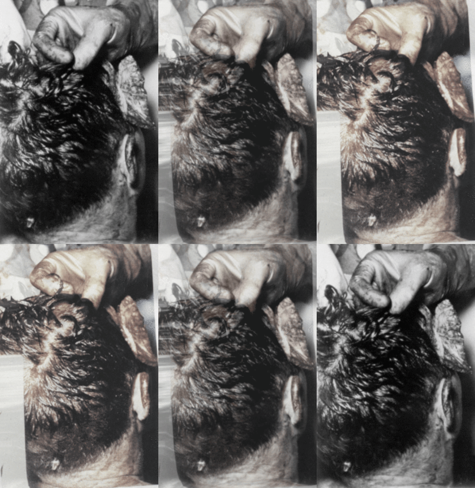
Hogwash. Dr. Mantik studied F3 (the back-of-the-head photo) with a stereo viewer at the National Archives and found that it does *not* show stereoscopic consistency, which means it has been faked.

Sorry Griffith but your continued claims that any photos/film which doesn't agree with your Loony Toons conspiracy is "faked" is becoming increasing tedious and not convincing anybody, so far you've got an army of alterationist's making unbelievably impossible photo realistic images and unfortunately for your credibility you haven't yet produced one image/film expert who in any way describes how this fakery was actually accomplished, let alone a demonstration of an actual altered stereoscopic image as an example, your claims are just that, claims based on delusion.

And talk about sticking your head in the sand and openly embracing the last century, you take the cake, the back of head animation requires millions of computer cycles which was unheard of when your "expert" made his self serving observation and the best comeback you've got is the obviously biased Mantik claiming that F3 doesn't show "stereoscopic consistency", unbelievable!

And who are you're going to believe, Mantik with an obvious axe to grind or your own lying eyes? The stereoscopic images when recombined can only show a 100% smooth rotation if each and every hair, skin cell, wrinkle, wound and overall shape displays the exact same depth mapping relatively in each original photo and to suggest that the following graphic is based on images which aren't "stereoscopically consistent" is a physical impossibility because any image alteration would not allow a smooth rotation and would have floating artefacts but as is clear this is not the case, you and your claim is totally bogus. You lose!


JohnM

Offline John Mytton

  • Hero Member
  • *****
  • Posts: 4267
So your "answer" to all the mutually corroborating Bethesda and Parkland descriptions of the large head wound is to nit-pick McClelland's verbiage and to once again post your goofy top-of-head GIF? That's it?
Oh, boy.

So you're going to latch onto a hyper-literal interpretation of "look down" and simply ignore the fact that McClelland's description of the large head wound closely corresponds to nearly all the other descriptions of the wound? It is amazing to see the difference in how you treat witnesses like McClelland and how you treat witnesses like Brennan.


Goofy?, you are so far out of your depth that your constant ad homs are your only defence of your complete inability to grasp even the most basic of concepts. As explained in the last post the only way to create a smooth rotation is if all the information which amounts to thousands and thousands of pixels in each stereoscopic image have precisely identical depth mapping in each photo relative to the camera lens and again any anomalies would create floating artefacts but unfortunately for you and your kooky assertions we only see smooth rotation meaning that the originally photographed object possesses solid unaltered dimensions.


And as for McClelland's claims, in addition to looking down into a wound which could only be observed if JFK's head was as seen in the officially authenticated autopsy photos, how the heck could he make an accurate observation of a third of Kennedy's brain missing if he only looked into a hole that was a sliver from the side, the only way he could make this determination is if he saw a massive amount of missing skull which exposed the brains intact left hemisphere and the missing brain tissue from the right side as seen in the above animation, to deny this logical deduction is to deny the very fabric of existence.


JohnM

JFK Assassination Forum


Offline John Mytton

  • Hero Member
  • *****
  • Posts: 4267
Mr. SPECTER. Did you observe the condition of the back of the
     President's head ?
Dr. McCLELLAND. Well, partially; not, of course, as I say, we did
     not lift his head up since it was so greatly damaged. We
     attempted to avoid moving him any more than it was absolutely
     necessary, but I could see, of course, all the extent of the wound.

McClelland seems to say he could not see the very rear of the President's head.

    "That there was not only a horrible gaping wound but that it was
     a cavity that extended down into the head. And as I stood there
     holding the retractor, I was looking down into it all the time. I was
     no more than eighteen inches away from the wound all the time,
     standing just above it, which was ten to fifteen minutes at least."
          -- Robert McClelland, 1991

How can McClelland be "looking down" into a gaping wound at the back of the head if he's "standing just above it" such that he could see it "extended down into the head"? The President's head have to be turned so that the back of the head was upright.

    "He was in terrible shape; the right side of his brain had been
     blown out."
          -- Robert McClelland, 1993


JohnM

Offline Michael T. Griffith

  • Hero Member
  • *****
  • Posts: 929
Mr. SPECTER. Did you observe the condition of the back of the
     President's head ?
Dr. McCLELLAND. Well, partially; not, of course, as I say, we did
     not lift his head up since it was so greatly damaged. We
     attempted to avoid moving him any more than it was absolutely
     necessary, but I could see, of course, all the extent of the wound.

McClelland seems to say he could not see the very rear of the President's head.

    "That there was not only a horrible gaping wound but that it was
     a cavity that extended down into the head. And as I stood there
     holding the retractor, I was looking down into it all the time. I was
     no more than eighteen inches away from the wound all the time,
     standing just above it, which was ten to fifteen minutes at least."
          -- Robert McClelland, 1991

How can McClelland be "looking down" into a gaping wound at the back of the head if he's "standing just above it" such that he could see it "extended down into the head"? The President's head have to be turned so that the back of the head was upright.

    "He was in terrible shape; the right side of his brain had been
     blown out."
          -- Robert McClelland, 1993

So even in the statements you quote, McClelland said "I could see, of course, all the extent of the wound." And, needless to say, once JFK was pronounced dead, the doctors in the room had plenty of time to observe JFK's wounds, especially when Father Huber conducted the last rites and then when the nurses began to wash the body, pack the rear head wound with gauze squares, and wrap the head and body in sheets in preparation for placing the body in the casket.

I am still waiting for you guys to deal with the fact that McClelland's WC description of the large head wound and his later drawing of the wound closely agree with the descriptions and diagrams of the wound given/drawn by numerous other witnesses, including those given/drawn by the nurse who packed the large head wound with gauze, by the Secret Service agent who saw the wound for several minutes up-close on the way to the hospital and then again at Bethesda, by the nurse who held a pressure dressing against the wound, by the mortician who reassembled the skull after the autopsy, and by the mortician's assistants who helped him prepare the body for burial, not to mention the descriptions in the 11/22/63 Parkland medical reports.

No rational person is going to buy the "they were all mistaken" argument. That dog just won't hunt when you're talking about multiple witnesses in three different locations who saw the wound up-close, who held the head, who packed the wound, etc., especially when many of them were unaware that others had given similar descriptions.

Finally, regarding Mytton's goofy top-of-head GIF, I repeat the fact that Tom Robinson, the mortician, explained to the ARRB that the top of JFK's head had no visible damage until Humes took a saw to it, and that the damage seen in the top-of-head photos was done by Humes and Boswell before the 8:00 PM autopsy started.

« Last Edit: August 14, 2020, 04:44:19 PM by Michael T. Griffith »

Offline John Iacoletti

  • Hero Member
  • *****
  • Posts: 10812


Typical "Mytton".  Sticks an arrow in his preferred location and ignores what the guy's hand is actually doing.

Just like it's typical "Mytton" to claim that a computer generated "rotation" somehow demonstrates anything about the underlying photographs.

JFK Assassination Forum